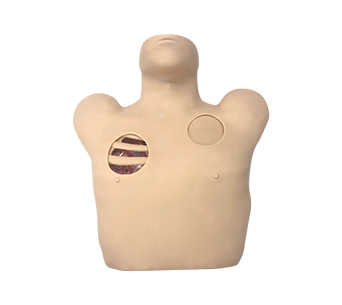
■ The simulator is the upper body of an adult, with a realistic shape and texture, and a standard puncture position.
■ The anatomical landmarks are obvious and can touch the clavicle, sternal incisions, ribs, intercostal spaces, etc., making it easy to operate and locate.
■ The simulator can be used for training closed drainage operations for pneumothorax and hydrochorax after chest trauma, as well as postoperative care for drainage tubes.
■ There are two "windows" on the left chest, located between the 2nd and 3rd ribs of the midclavicle and the 5th and 8th ribs of the anterior axillary line. The windows can display the anatomical structures of the chest wall, including muscles, blood vessels, nerves, etc., for easy teaching and operation.
■ On the right is the operating area, which allows for thoracic puncture and closed thoracic drainage operations, as well as postoperative nursing exercises for drainage tubes. When entering the chest, there is a clear sense of emptiness.
■ Correct puncture can expel pleural effusion/gas.
■ Simulating pleural effusion can adjust the color and viscosity of lesions to different degrees for disease diagnosis.
■ The skin and puncture module can be repeatedly punctured and easily replaced.


