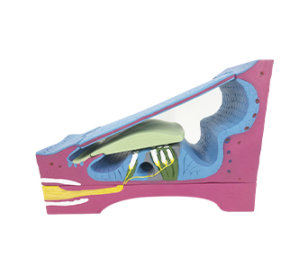
■ The spiral apparatus and membranous cochlear canal simulator can be enlarged 350 times and divided into 5 parts, displaying the three-dimensional microstructure of the three walls of the spiral apparatus and membranous cochlear canal. The inner end of the simulator is a bony spiral plate, which is equivalent to the cross-section at the spiral edge. The bone, the thickened periosteum on the surface, and the auditory nerve fiber bundle penetrating through the bone can be seen. The other end of the simulator is a spiral ligament, which contains most blood vessels. From a side view, the vestibular membrane can be seen rising from the periosteum above the spiral edge, Stop above the spiral ligament. When the vestibular membrane is removed for observation, it can be seen that it is composed of the upper mesothelium, the middle connective tissue, and the lower epithelium. The outer wall of the membranous cochlear duct is a spiral ligament, with a single layer of cuboidal epithelium attached to the inner surface.
■ Size: 350 times enlarged, 47.5 x 18 x 32.5cm
■ Material: PVC


