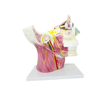
■ The simulator has a partial skull base, and the upper and outer walls of the orbit have been dissected, exposing the eyeball, extraocular muscles, intraorbital nerves, mandibular branches, as well as some temporal bones and superficial neck muscles. The distribution of brain nerves in the neck has been exposed; Twelve pairs of cranial nerves can be seen entering and exiting the cranial foramen fissure on the inner surface of the skull base. In the middle plane, olfactory nerve fibers can be seen passing through the ethmoidal foramen and entering the olfactory bulb in the mucosa of the upper turbinate. The lateral wall of the nasal cavity is incised to expose the pterygopalatine ganglion and its branches;The distribution of the positions of the eyeball, extraocular muscles, oculomotor nerve, trochlear nerve, abductor nerve, trigeminal nerve ophthalmic branch, and ciliary ganglion in the orbit;The mandibular branch and part of the temporal bone have been cut off, revealing the maxillary nerve of the trigeminal nerve, the course, branches, distribution, and inferior mandibular ganglion of the mandibular nerve;The facial nerve divides into the superficial petrosal nerve and the chordae tympani within the facial nerve canal, the glossopharyngeal nerve and vagus nerve run and distribute in the neck, the accessory nerve innervates the sternocleidomastoid and trapezius muscles, and the hypoglossal nerve enters the tongue.
■ Size: 2x enlarged, 42.5 x 15 x 40cm
■ Material: PVC


