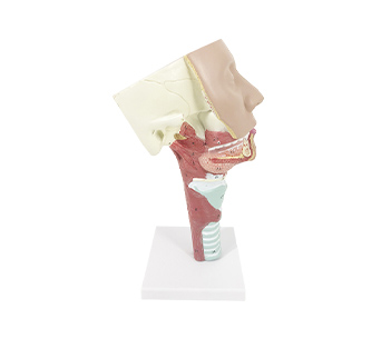
■ There are anterior and middle cranial fossa with a skull base. The front is the face, the lower chin, and the throat and trachea in front of the neck, and the back is the posterior wall of the pharynx and upper esophagus. From the back of the simulator, all pharyngeal constrictors and their attachments can be seen. The pharyngeal constrictor muscle originates from the buccal pharyngeal suture, the pharyngeal constrictor muscle originates from the greater angle of the hyoid bone, and the pharyngeal constrictor muscle originates from the oblique line of the thyroid cartilage, all of which stop at the pharyngeal suture. The three muscles are arranged in a layered pattern from bottom to top, From both sides, the relationship between the styloid process pharyngeal muscle, styloid process lingual muscle, and styloid process hyoid muscle can be seen, as well as the positional relationship between the pharynx and larynx, esophagus and trachea.
■ The three major salivary glands fully expose their positions and the direction of their ducts. The parotid gland is located on the surface of the posterior mandibular fossa and the posterior edge of the masseter muscle. The parotid gland duct crosses the masseter muscle below the zygomatic arch to reach its anterior edge deep into the cheek. The submandibular gland duct originates from the deep part and enters the deep surface of the sublingual gland
■ The sublingual gland is located above the jawbone muscle, and the sublingual gland duct opens at the mucosa of the tongue floor.


